



Researchers from the COR group at the Institute of Information and Communications Technologies (ITACA) of the Universitat Politècnica de València (UPV) have developed an artificial intelligence model that can locate and estimate the extent of tissue abnormalities associated with atrial cardiomyopathy using electrical recordings from the body’s surface.
The system, based on graph neural networks (GNNs), achieved 89% accuracy in locating affected areas and 84% accuracy in determining the extent of damage to the atrial tissue. It also retained this capability when analysing anatomies that had not been used during training.
The next step: validation in patients
The work remains a proof of concept based on simulated data, so its results do not support immediate clinical use. The next step will be to validate the model using recordings from real patients. As the study itself notes, “prospective validation with clinical recordings is needed before this approach can be translated into treatment-planning applications”.
If the results are confirmed in clinical studies, this technology could help pave the way for new tools to characterise atrial tissue without invasive procedures, while providing further information to support the study and treatment of abnormalities associated with atrial cardiomyopathy.
Published in the scientific journal Discover Computing, the study was led by María Macarulla-Rodríguez, a researcher in COR-ITACA. It also involves Jorge Sánchez, Andreu M. Climent and María S. Guillem, researchers at ITACA and members of Corify Care S.L.; Cristian Barrios Espinosa and Axel Loewe (Karlsruhe Institute of Technology); and Ernesto Zacur (Corify Care S.L.).
“The study’s main finding is that combining body-surface electrical maps, spatio-temporal analysis and graph neural networks has considerable potential for the non-invasive characterisation of atrial cardiomyopathy”, says María Macarulla, lead author of the study.
A new way to study atrial tissue
Atrial cardiomyopathy encompasses electrical and structural abnormalities in atrial tissue, such as fibrosis, which are associated with the onset and progression of atrial fibrillation, one of the most common cardiac arrhythmias.
“Knowing where the affected tissue is located and how extensive it is could help us better understand the disease and, potentially, plan treatments such as ablation», explains María S. Guillem, director of ITACA and a contributor to the study.
These abnormalities can currently be characterised using techniques such as invasive intracardiac electroanatomical mapping or certain magnetic resonance imaging tests.
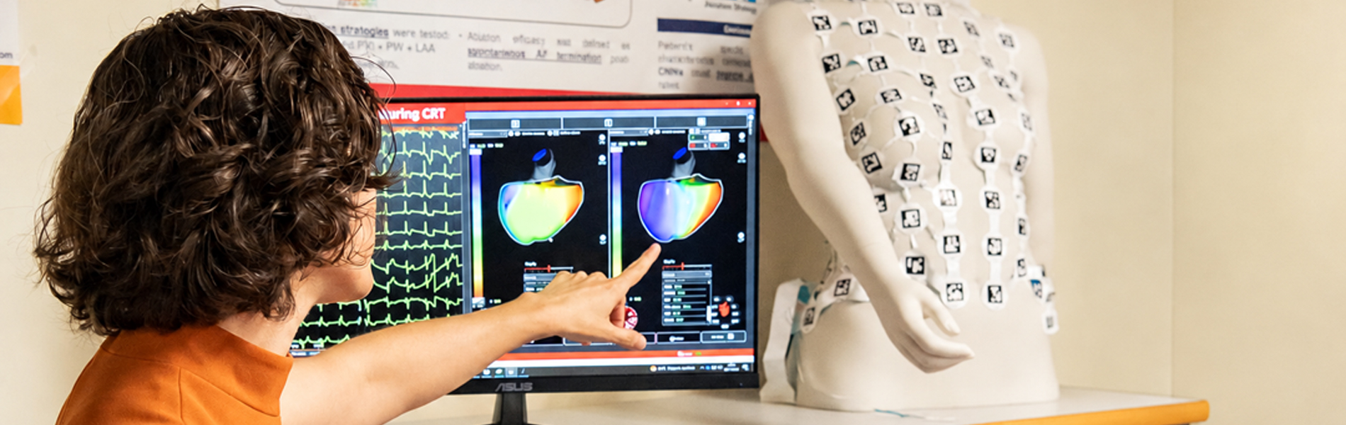
The new approach uses body-surface potential maps (BSPMs), obtained by placing electrodes across the torso to record the heart’s electrical activity.
“The artificial intelligence then analyses how these signals are distributed across space and how they change over time, to identify patterns associated with the location and extent of the affected tissue,” explains the ITACA director.
89% accuracy in locating affected tissue
To develop and evaluate the model, the team used 14,400 simulated body-surface potential maps, generated from different atrial and torso anatomies and covering different degrees and locations of atrial cardiomyopathy.
“In the reference configuration, with 128 electrodes, the model achieved 89% accuracy in locating the affected tissue, with a mean sensitivity of 89% and a mean specificity of 90%”,, highlights María Macarulla.
One of the most significant results was that the system maintained good performance when analysing anatomies that differed from those used to train it. This is an important consideration for any future application in people with different anatomical characteristics. “The model also achieved an overall accuracy of 84% in determining the extent of the affected tissue. We also found that its performance remained relatively stable when signal quality decreased, suggesting a degree of robustness to noise,” says María Macarulla.
The results also show that the anatomical diversity of the training data is crucial: including a wider variety of atrial and torso models improved the system’s classification performance.
Reference: A graph neural network framework for characterizing atrial cardiomyopathy from body surface potential maps. María Macarulla-Rodríguez, Jorge Sánchez, Cristian Barrios Espinosa, Axel Loewe, Ernesto Zacur, Andreu M. Climent & María S. Guillem. https://tinyurl.com/yc8ye2z3