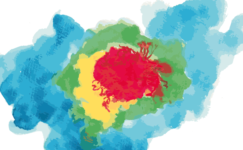
CURIAM BT+ is a computational system designed to assist experts in identifying tissue signatures associated with angiogenesis and vasculogenesis in primary glioblastoma tissues. It aims to predict tumour aggressiveness, patient survival and facilitate personalised monitoring of this complex condition. This tool improves surgical and treatment planning, recurrence detection, and survival prediction by providing an insightful, reproducible nosology map of each patient’s glioblastoma tissue.
Applications
- Clinical Oncology: For clinicians treating glioblastoma, CURIAM BT+ can aid in assessing tumor aggressiveness, predict patient survival, and monitor the disease over time.
- Radiology: Radiologists could use CURIAM BT+ to improve the interpretation of medical imaging scans related to glioblastoma, enabling more accurate assessments.
- Neurosurgery: For neurosurgeons, CURIAM BT+ could be used to guide surgical planning and treatment strategy by offering a detailed map of the tumor and surrounding tissues.
- Research: Scientists studying glioblastoma could use CURIAM BT+ to gain insights into tumor behavior, aiding in the development of new treatments and interventions.
- Pharmaceuticals: Drug developers could potentially use CURIAM BT+ to evaluate the effectiveness of new drugs and treatments for glioblastoma.
Technology Overview

Glioblastoma is the most frequent type of aggressive central nervous system tumour. The segmentation and analysis of tissues related to this lesion, such as viable tumours, oedema or necrosis, and the classification of different sub-types of tissues derived from these regions, is crucial to follow the evolution of tumour growth during therapy.
CURIAM BT+ is a computational system that allows experts to intuitively observe tissue signatures related to angiogenesis and vasculogenesis in primary glioblastoma tissues and predict the aggressiveness of these tumours and the expected survival of the patient. This system provides a tool to promote personalised monitoring of this complex pathology. The segmentation of tissue signatures in the viable tumour, in the peritumoral area and the oedema provides the necessary knowledge for better surgical and treatment planning, recurrence detection and survival prediction.
CURIAM BT+ generates a customised and reproducible nosology map of the patient where multi-parametric tissue signatures describe each glioblastoma tissue. CURIAM BT+ performs a non-supervised multi-parametric classification based on structured prediction algorithms, which improve the consistency and reproducibility of the nosology maps, and allow the inclusion of spatial/temporal information from available longitudinal studies of the patient to respond to the need for information requested from clinical settings for better patient follow-up.


