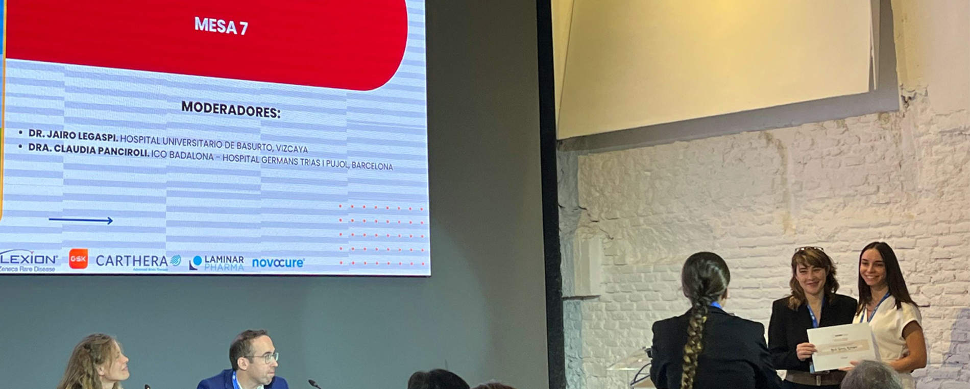
The Biomedical Data Science Lab (BDSLab) of the ITACA Institute at the Universitat Politècnica de València (UPV) has received the Best Oral Presentation Award at the XVII Symposium of the Grupo Español de Investigación en Neurooncología (GEINO), held in Madrid on 28–29 October.
The award-winning work, presented by María Gómez Mahiques, proposes an automatic and reproducible methodology for the delineation of the non-contrast-enhancing tumour (nCET) in IDH wild-type glioblastomas — one of the most aggressive and poor-prognosis types of brain tumours.
The study, conducted by researchers from BDSLab-ITACA under the supervision of Elies Fuster-García and Juan M. García-Gómez, addresses one of the main challenges in current neuroimaging: accurately identifying tumoural areas that do not show enhancement on conventional magnetic resonance imaging, which reflects tumour infiltration processes.
“This region, invisible in traditional images, may contain infiltrative tumour tissue. Its identification is key to understanding the true extent of the tumour and improving treatment planning, such as determining the area to be removed during neurosurgery,” explains María Gómez Mahiques, researcher at the ITACA Institute.

Artificial intelligence to enhance diagnostic and therapeutic precision
The methodology developed represents a significant advance in the application of artificial intelligence to medical imaging, enabling objective, automatic, and reproducible delineation of regions that previously relied on manual interpretation by specialists.
According to BDSLab researchers, a more precise identification of the nCET could improve the characterisation of tumour infiltration and provide prognostic value, with a direct clinical impact on surgical and radiotherapy planning.
“This methodology helps to make more personalised and data-driven decisions”, conclude the authors of the study.
Noticia elaborada por ITACA FORWARD, financiada por Ivace+i Innovación y la Unión Europea a través del Programa Fondo Europeo de Desarrollo Regional (FEDER) Comunitat Valenciana 2021-2027 (Referencia del proyecto: INNVA2/2025/17)


